


U.S. East Coast hospital
Hospital adopts an enterprise imaging strategy to integrate point-of-care images into the EMR.
Hospital adopts an enterprise imaging strategy to integrate point-of-care images into the EMR.

Historically, medical imaging was almost exclusively a function of the Radiology and Cardiology departments within a healthcare system. However, the rise of specialty imaging devices, portable modalities, smartphones and other mobile devices has increasingly extended imaging outside of Radiology and Cardiology and to the point of care. This growing trend has created a number of imaging silos within healthcare organizations that are disconnected from core clinical systems or patient records. The result is scattered medical information that is largely invisible or inaccessible to clinicians, which can negatively impact diagnosis, treatment and care.
This hospital realized how important these point-of-care images are to clinical outcomes and recently embarked on an initiative to consolidate and manage these assets at an enterprise level.
In 2013, the hospital began an enterprise imaging assessment to identify and understand the needs of all the imaging-intensive departments within the organization. This included not just Radiology and Cardiology areas but all the “ologies” including the Dermatology, Ophthalmology, Pathology departments and more.
The goal was to gain awareness of all imaging workflows and recommend an action plan that would support a unified image management approach for all departments that leveraged as much of the hospital’s existing infrastructure as possible. The hospital also wanted to ensure this strategy included the management of video clinical assets — a largely overlooked aspect of many imaging initiatives.
An enterprise imaging steering committee was established that included a mix of clinical, administrative and IS personnel. It was led by the PACS/Radiology informatics manager and IS strategy leaders with previous governance expertise. The committee identified 43 imaging-intensive clinical departments and conducted interviews with the clinical and administrative leaders of each to determine their existing imaging workflows, processes and storage policies.
Much was learned during this discovery exercise — from both a radiology and point-of-care imaging perspective. For instance, the hospital discovered that it had 21 different image acquisition and viewing solutions (e.g. PACS viewer, ultrasound application viewer, image sharing application viewer, enterprise viewer, etc.) in use throughout the hospital and 10 disparate image storage solutions.
“There was a lot of storage we knew about, including our Acuo vendor neutral archive (VNA), the OnBase repository and the Epic BLOB server,” their radiology informatics manager said. “But, there was also a lot of storage we didn’t have insight into such as memory cards, shared drives, local hard drives and smartphones with cloud storage. This was a concern from both a clinical and data security standpoint.”
For the most part, radiology and cardiology DICOM modalities (e.g. MRI, CT, etc.) were all stored in their existing Acuo VNA and were viewed using the radiology PACS or the enterprise viewer via its Epic EMR. However, the provider was surprised to learn about the growth in image sharing of its DICOM assets.
“The number of departments using image sharing for DICOM images increased significantly in just a few years,” says the radiology informatics manager. “Inbound DICOM image sharing was typically handled using our image sharing application server while outbound DICOM sharing was conducted by burning CDs or DVDs or using the requesting facility’s image sharing application. This created multiple places where DICOM images were stored.”
When it came to non-DICOM images and video (i.e. JPEG, TIFF, PDF, AVI, MPG, etc.) things got more complicated. For example, the hospital discovered that while most attending physicians used Epic Haiku to view non-DICOM images, trainees, residents and fellows mainly used their personal smartphones.
In most instances, these physicians didn’t even know that Haiku existed or weren’t provided a hospital issued mobile device with Haiku preloaded.
The hospital also identified numerous third-party applications, scopes and cameras that were used for non-DICOM image and video capture. Areas such as the ER needed to capture images on smartphones because this was the quickest way to document patient conditions in some emergency situations (i.e. patient seizures, stroke, trauma, etc.).
These non-DICOM images and videos came in multiple formats and were stored in a wide variety of locations including the Epic Media Tab in Haiku, hospital shared network drives, individual or department hard drives, specialty imaging/video systems (e.g. OR and ENT scopes), smartphone storage, cloud storage and camera memory cards. In many instances, images and video were left on a device with no defined storage policy and no association or tie-in to a specific patient identifier in the EMR.
A lack of integration between non-DICOM capture devices and Epic also forced some physicians to implement a workaround where they emailed themselves in order to get an image to the network drive so they could upload it onto Epic’s Media Tab. An initiative in June 2015 showed that 6,095 images were uploaded to Epic using personal email, from which they were cut and pasted into a note.
Radiology informatics manager US East Coast Hospital
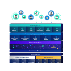
While the hospital realized no single solution would solve all the clinical imaging and video needs of the organization, it did believe the best approach was to implement multiple solutions from a single vendor to achieve its desired outcome. That vendor was Hyland Healthcare.
They already leveraged Acuo as the primary storage layer for all DICOM images from Radiology and Cardiology. The decision was made to enable the VNA to support the nonDICOM images and video in play throughout the enterprise, too. This required the hospital to add XDS (cross-enterprise document sharing) and other functionality to its VNA.
The hospital also elected to implement NilRead as its new enterprise viewer. This viewer could display all clinical images (both DICOM and non-DICOM) and video in context of the patient record in Epic. The solution also offers advanced visualization capabilities that allow it to be used for diagnostic interpretation in some specialty imaging departments
Other aspects of the initiative included leveraging PACSgear Encounter Worklist to support non-DICOM order workflows (e.g. point-of-care ultrasound, GI lab, ophthalmology, etc.) and OnBase, Hyland’s enterprise information platform, and OnBase Patient Window to more easily connect images, video and other content to Epic’s Media Tab.
The hospitals go-forward strategy is relatively simple. Images and video of the patient (e.g. MRIs, CT scans, wound photos, surgical video, dermatology photos, etc.) are stored in Acuo. Images about the patient (e.g. driver’s license and insurance card scans) are stored in OnBase. This consolidation helps eliminate many of the imaging silos that previously existed at the hospital, simplifying image and video management throughout the enterprise.
“Bringing all clinical imaging together is best for our clinicians and patients,” the Radiology Informatics manager. “Our physicians will now know where all images are located and can access these assets in a single application. Prior to our enterprise imaging effort, we had a lot of imaging silos that our physicians couldn’t access or didn’t know existed. An enterprise imaging approach will allow us to provide better care to our patients because our physicians will have all the health information they need to make the best clinical decisions at their fingertips.”