University medical center
Healthcare organization gives faster, more complete answers for patients awaiting stroke diagnosis.

Harness the power of a unified content, process and application intelligence platform to unlock the value of enterprise content.
Learn more
Automate your document-centric processes with AI-powered document capture, separation, classification, extraction and enrichment.
Learn about Hyland IDPIt's your unique digital evolution … but you don't have to face it alone. We understand the landscape of your industry and the unique needs of the people you serve.
 Overview of industries
Countless teams and departments have transformed the way they work in accounting, HR, legal and more with Hyland solutions.
 Overview of departments
We are committed to helping you maximize your technology investment so you can best serve your customers.
 Overview of services

Discover why Hyland is trusted by thousands of organizations worldwide.
Hear from our customers
Our exclusive partner programs combine our strengths with yours to create better experiences through content services.
Overview of partners
Join The Shift newsletter for the latest strategies and expert tips from industry leaders. Discover actionable steps to stay innovative.
Register now
Hyland connects your content and systems so you can forge stronger connections with the people who matter most.
Learn about HylandWith our modern, open and cloud-native platforms, you can build strong connections and keep evolving.
 Dig deeper
Reading time minutes
Healthcare organization gives faster, more complete answers for patients awaiting stroke diagnosis.

Telemedicine is a vital component in delivering effective healthcare — particularly to those in outlying areas. Telemedicine also offers opportunities to capture efficiencies in collaborative workflows for viewing medical imaging.
When one university medical center wanted to find ways to be more effective in delivering care to possible stroke patients in outlying areas, it went in search of a solution that would support viewing and collaborating on images more easily, regardless of location.
The university was looking for solutions to better support televiewing, but they were also looking to resolve some other standing challenge.
Traditional PACS experience heavy traffic from the pre-fetching of prior studies and other non-diagnostic viewing requests. To reduce that traffic burden and make it easier for clinicians and staff to share images and collaborate, the university wanted to adopt a zero-footprint universal enterprise diagnostic image viewer. This would also free up IT staff from managing local client installs — which can be particularly challenging when supporting outlying communities.
Like many others that serve their communities, this university medical center received tens of thousands of patient CDs every year from hundreds of outside facilities. Clinicians were forced to wait as images were manually imported from the CDs — and then wait for patient demographics to be manually matched. Using VPNs as an alternative approach to the CD-sharing process created bottlenecks and incurred significant overhead. As these delays could delay patient care, a better approach was needed.
Medical researchers in the university wanted the ability to upload, access and share imaging studies and other data while also segregating this information from active patient data and content. Researchers would sometimes turn to freeware applications that were not designed for this purpose and also posed security risks. The freeware tools were mostly client-server applications that tied a researcher to a specific workstation and prohibited access to images in other locations or devices.
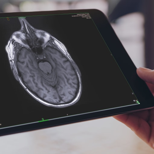
The medical center moved its enterprise users to NilRead, the Hyland Healthcare zero-footprint enterprise diagnostic viewer, with the NilFeed extension that allows a secure connection using SSL and https without a VPN. This provided efficiencies for faster access and viewing of images.
This change dramatically reduced the traffic workload on the main PACS — which would be primarily used for diagnostic reads within radiology department. University physicians gained the flexibility of a web-based viewer that allowed them to view images simultaneously and use collaboration tools from any location. The change also freed IT staff from managing local client installs.
The ability to ingest images using NilRead allows the images to be stored directly in the VNA. With this functionality, the university was able to directly transfer patient images from referring institutions instead of waiting for CDs to be produced, transported and ingested into the university system.
While CDs have not been completely eliminated from all locations, the workload from high-volume CD senders was significantly reduced. This reduced work queues for imaging library staff and sped up the turnaround time for CDs sent to the department.
Now, clinical researchers from outside the university could easily send imaging data, streamlining the process. Storing images in Hyland Healthcare’s Acuo VNA also provided sophisticated tools that allowed the research imaging data to be segregated from live patient data.
The university gained more than a robust, diagnostic-quality viewer — they gained the flexibility to access images from remote locations and portable devices. This, in turn, allowed them to create a teleneurology program to serve the outlying areas.
Among the most innovative uses of NilRead’s power is managing urgent neurology consults from a remote location — in other words, teleneurology. Since the start of the teleneurology program, patients around the region gained faster access to specialists for stroke diagnoses. Patients deemed not to be experiencing a stroke no longer had to be transported to the university hospital for further care and assessment. This freed up beds, eliminated travel demands on patients and their families and decreased unnecessary testing on patients. By the same token, patients that were indeed experiencing a stroke per the collaborative consults were sent immediately to the university for care. The teleneurology process also saves time on the stroke protocol pathway for patients upon their arrival.
The referring facilities can now treat patients locally, keeping them close to family and familiar caregivers while preserving beds for patients with severe cases.
By implementing a telestroke program, patients can receive faster access to specialists for diagnoses, which greatly impacts patient outcomes. With the success of the teleneurology program, telemedicine capabilities have been extended to other service lines through enhanced image sharing.